
Pemphigus vulgaris is a life-threatening autoimmune blistering disease of the skin and mucous membranes. Often misdiagnosed initially, its rapid progression and painful lesions make early recognition and treatment crucial.
In this article, we will cover its pathophysiology, diagnosis, clinical findings, and treatment approach, based on real-world emergency and clinical dermatology practice.
🧬 What is Pemphigus Vulgaris?
Pemphigus vulgaris is an autoimmune disorder characterized by the destruction of desmosomes, the intercellular structures that hold epidermal skin cells (keratinocytes) together.
🧪 Pathophysiology in Brief:
- Desmosomes = “Glue” between epidermal cells
- Autoantibodies (IgG) are formed against desmoglein-3 (and sometimes desmoglein-1)
- This causes acantholysis — the loss of cohesion between keratinocytes
- Result: Intraepidermal blistering and flaccid bullae
🧫 Histology & Immunofluorescence
🔍 Biopsy (Gold Standard Test):
- Shows intraepidermal clefting
- Acantholytic cells seen floating in blister cavity
- Loss of cohesion among keratinocytes
🌟 Direct Immunofluorescence (DIF):
- Intercellular IgG deposits in a “fishnet” pattern around keratinocytes
- Diagnostic hallmark of pemphigus vulgaris
🧍♀️ Case Presentation: Classic Clinical Scenario
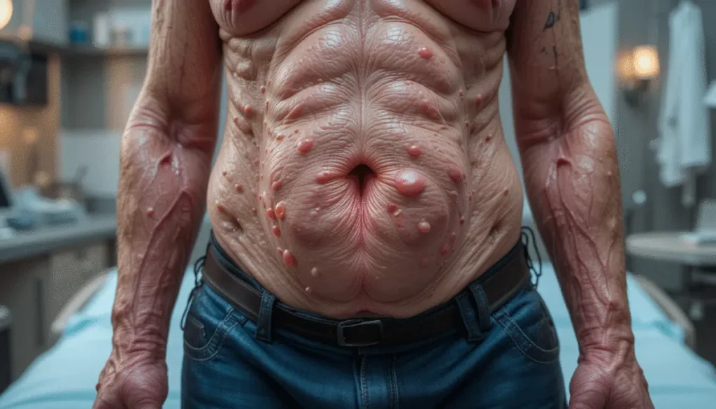
A 38-year-old woman presents with painful fluid-filled lesions over face, limbs, and oral mucosa. Lesions started with facial erythema and rapidly spread. She reports severe pain but no itching. Blisters rupture easily, leading to erosions and raw denuded skin.
🧠 Key Clinical Features:
| Feature | Description |
|---|---|
| Blisters | Flaccid, easily rupturing |
| Nikolsky Sign | Positive: Skin peels off with mild friction |
| Oral Involvement | Common, painful erosions |
| Pain | Severe, out of proportion |
| Pruritus (Itching) | Usually absent |
| Systemic symptoms | Dehydration, secondary infections |
❗ Differentiation from Bullous Pemphigoid
| Feature | Pemphigus Vulgaris | Bullous Pemphigoid |
|---|---|---|
| Blister location | Intraepidermal | Subepidermal |
| Oral involvement | Common | Rare |
| Nikolsky sign | Positive | Negative |
| Blister type | Flaccid, easily ruptured | Tense, less likely to rupture |
| Autoantibodies | Anti-desmoglein 3 | Anti-hemidesmosome (BP180, BP230) |
💉 Diagnosis
- Clinical signs: Flaccid bullae, oral ulcers, positive Nikolsky sign
- Skin biopsy – H&E staining shows acantholysis
- Direct immunofluorescence (DIF) – “Fishnet” pattern
- ELISA – Anti-desmoglein 3 and 1 antibodies
💊 Treatment Plan
Pemphigus vulgaris is a dermatologic emergency. Untreated, it can lead to sepsis, electrolyte imbalance, and death.
🔹 First-line:
- High-dose systemic corticosteroids (e.g. Prednisone)
- Supportive care: IV fluids, wound care, pain control, nutritional support
🔹 Add-on or steroid-sparing agents (if needed):
- Azathioprine
- Mycophenolate mofetil
- Cyclophosphamide (rare)
🔹 Severe/refractory cases:
- IVIG (Intravenous Immunoglobulin) — neutralizes pathogenic autoantibodies
- Rituximab — anti-CD20 monoclonal antibody, depletes B cells
🚑 Emergency Supportive Measures
Treat just like a burn patient:
| Support | Details |
|---|---|
| IV fluids | Replace volume lost from skin barrier damage |
| Analgesia | Morphine or NSAIDs for severe pain |
| Antibiotics | Prevent/treat sepsis from open erosions |
| Wound care | Sterile dressings, avoid trauma |
| Nutrition | Oral mucosal lesions may impair intake |
🧠 In Summary
| Concept | Key Takeaway |
|---|---|
| Disease Mechanism | Autoantibodies destroy desmosomes |
| Diagnostic Tests | Biopsy, Immunofluorescence (fishnet pattern) |
| Clinical Hallmarks | Flaccid blisters, mucosal erosions, painful |
| Nikolsky Sign | Positive |
| Treatment | Systemic steroids → Immunomodulators → IVIG/Rituximab |
| Supportive Care | Manage like burns: fluids, antibiotics, pain |