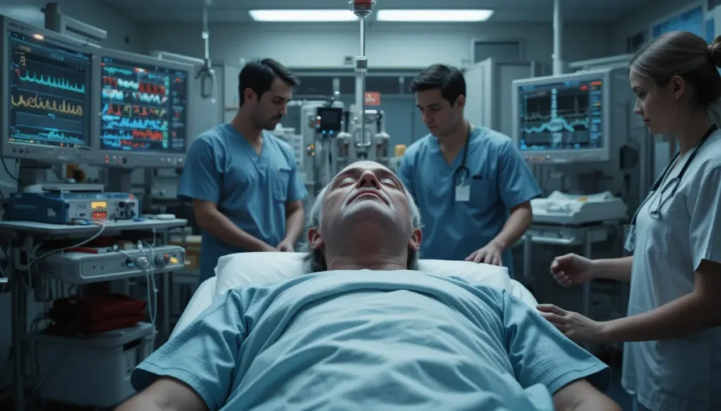
Cardiac arrest is a sudden, life-threatening event in which the heart stops pumping blood effectively. Without immediate intervention, it leads to irreversible brain damage and death within minutes. Understanding the correct approach to an unconscious, pulseless patient is critical in emergency care — whether you’re a healthcare professional or trained responder.
🫀 What is Cardiac Arrest?
Cardiac arrest is defined as the abrupt cessation of heart activity, resulting in:
- Loss of consciousness
- Absent carotid or major pulses
- No effective breathing
The most common scenario: A person suddenly collapses, becomes unresponsive, is not breathing, and has no pulse.
🔎 Causes of Cardiac Arrest
Cardiac arrest can result from various cardiac and non-cardiac conditions:
- Ventricular Fibrillation (VFib) – Disorganized electrical activity prevents the ventricles from pumping.
- Asystole – No electrical or mechanical heart activity (flatline).
- Pulseless Electrical Activity (PEA) – Electrical rhythm present on ECG, but no palpable pulse due to mechanical failure.
- Massive PE, cardiac tamponade, trauma, electrocution, MI, or drug toxicity
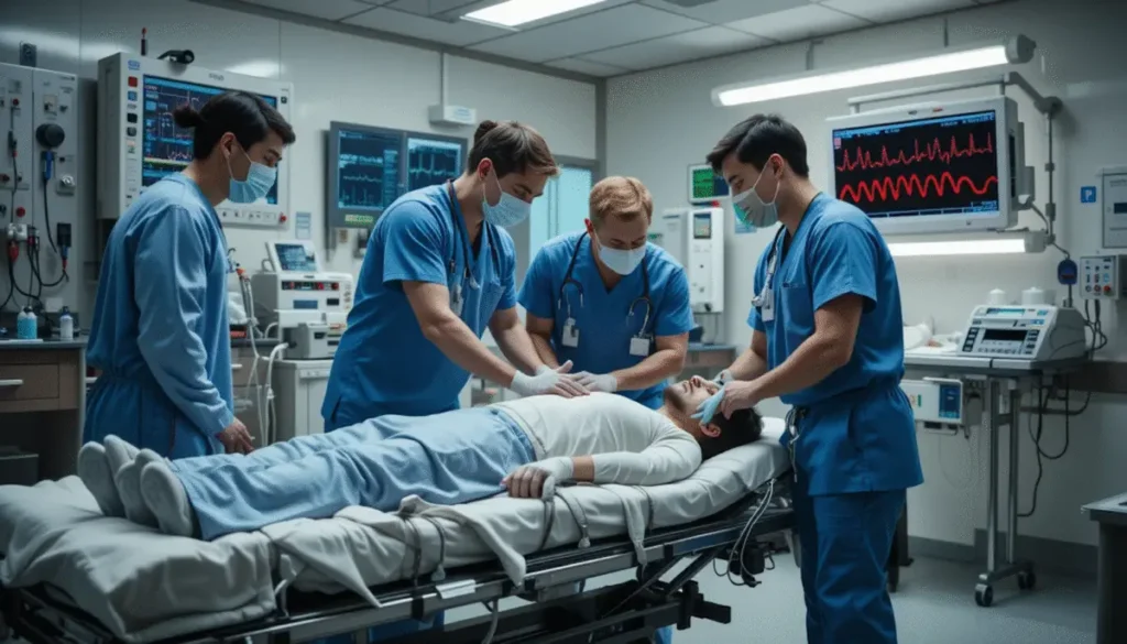
🚨 First Response to Cardiac Arrest
Key Signs:
- Unconsciousness
- No breathing
- No palpable carotid pulse
Immediate Actions:
- Call for help and activate emergency response.
- Place patient on a firm, flat surface, face up.
- Simultaneously check carotid pulse and breathing (no more than 10 seconds).
- If no pulse → begin high-quality CPR immediately.
🔁 CPR – Chest Compression Basics
- Rate: At least 100–120 compressions per minute
- Depth: At least 2 inches (5 cm)
- Compression-to-ventilation: 30:2
- Hand position: Lower half of sternum, elbows locked, shoulders directly over hands
- Allow full chest recoil between compressions
- Minimize interruptions
If outside a hospital, provide rescue breaths mouth-to-mouth. In-hospital, use a bag-valve mask or airway adjunct.
⚡ Shockable vs. Non-Shockable Rhythms (ACLS)
Once defibrillator and monitor are attached, rhythm determines next steps.
| Rhythm Type | Examples | Defibrillation? |
|---|---|---|
| Shockable | Ventricular Fibrillation (VF), Pulseless VTach | ✅ Yes |
| Non-Shockable | Asystole, Pulseless Electrical Activity (PEA) | ❌ No |
📋 ACLS Algorithm: Shockable Rhythm (VF / VT)
- Start CPR
- Attach monitor/defibrillator
- If shockable rhythm:
➤ Deliver shock (120–200J biphasic or AED auto dose)
➤ Resume CPR for 2 minutes
➤ Establish IV access
➤ Give epinephrine 1 mg IV every 3–5 minutes
➤ Reassess rhythm → if still shockable
➤ Deliver second shock
➤ Resume CPR
➤ Give amiodarone 300 mg IV, then 150 mg if needed
➤ Continue CPR + rhythm checks + further shocks
🛑 ACLS Algorithm: Non-Shockable Rhythm (Asystole / PEA)
- Start CPR immediately
- Establish IV access
- Give epinephrine 1 mg IV every 3–5 minutes
- Continue CPR
- Monitor ECG continuously
- Search for reversible causes (the “Hs and Ts”)
🧪 The Hs & Ts – Reversible Causes
| Hs | Ts |
|---|---|
| Hypovolemia | Tension pneumothorax |
| Hypoxia | Tamponade (cardiac) |
| Hydrogen ion (acidosis) | Toxins |
| Hypo-/Hyperkalemia | Thrombosis (MI or PE) |
| Hypoglycemia | Trauma |
| Hypothermia |
🫁 Post-Resuscitation Care
If the patient regains pulse (ROSC – Return of Spontaneous Circulation):
- Begin post-arrest management: oxygenation, blood pressure support, targeted temperature management
- Investigate and treat the underlying cause
- Transfer to intensive care immediately
✅ Summary
- CAB approach: Circulation, Airway, Breathing — prioritize compressions
- Shockable rhythms: VFib, pulseless VT → Defibrillate + epinephrine + amiodarone
- Non-shockable rhythms: Asystole, PEA → Epinephrine + CPR (no defibrillation)
- High-quality CPR and rhythm reassessment every 2 minutes
- Look for treatable causes to improve outcomes
Early, consistent, and accurate application of ACLS protocols can significantly improve survival in cardiac arrest patients.