Impetigo is one of the most common superficial bacterial skin infections, particularly affecting children. It is highly contagious and frequently seen in hot, humid climates, or overcrowded settings with poor hygiene. This post explores the pathophysiology, presentation, and step-by-step treatment approach to impetigo, including its two clinical types: non-bullous and bullous impetigo.
👶 Case Scenario
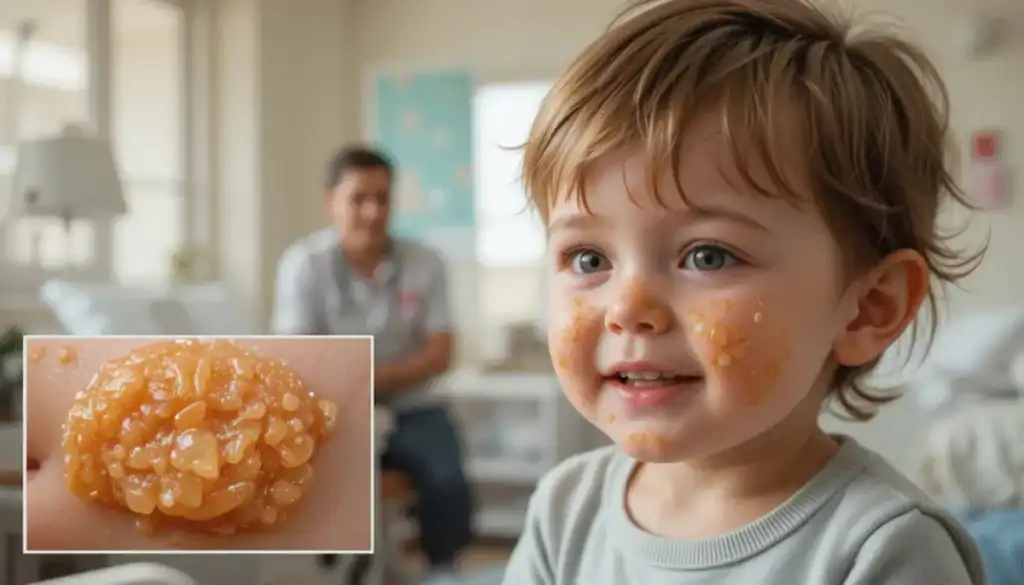
A 3-year-old boy from a rural background presents with thick, golden-yellow crusted lesions around the lips and mouth. He has no systemic illness, vitals are normal, and he is playful. The lesions are not painful, but mildly itchy. On examination, regional lymph nodes are palpable.
Diagnosis: Impetigo (Non-Bullous type)
🧬 What is Impetigo?
Impetigo is a highly contagious superficial infection of the skin involving only the epidermis. It is most commonly caused by:
- Staphylococcus aureus
- Group A beta-hemolytic Streptococcus (Streptococcus pyogenes)
🔍 Risk Factors:
- Poor hygiene
- Overcrowded environments (daycares, rural settings)
- Warm and humid weather
- Minor trauma or insect bites
🧪 Pathology Overview
The skin has three layers:
- Epidermis (superficial)
- Dermis (middle)
- Hypodermis (deep, fat and vessels)
Impetigo affects the epidermis, making it a non-scarring, superficial infection.
🔍 Types of Impetigo
1. Non-Bullous Impetigo (Most Common)
| Feature | Description |
|---|---|
| Cause | Streptococcus pyogenes ± S. aureus |
| Lesions | Honey-colored crusted erosions |
| Location | Around the mouth and nose |
| Symptoms | Painless, mildly itchy |
| Lymphadenopathy | Often present |
📸 Honey-crusted lesions are classic in non-bullous impetigo.
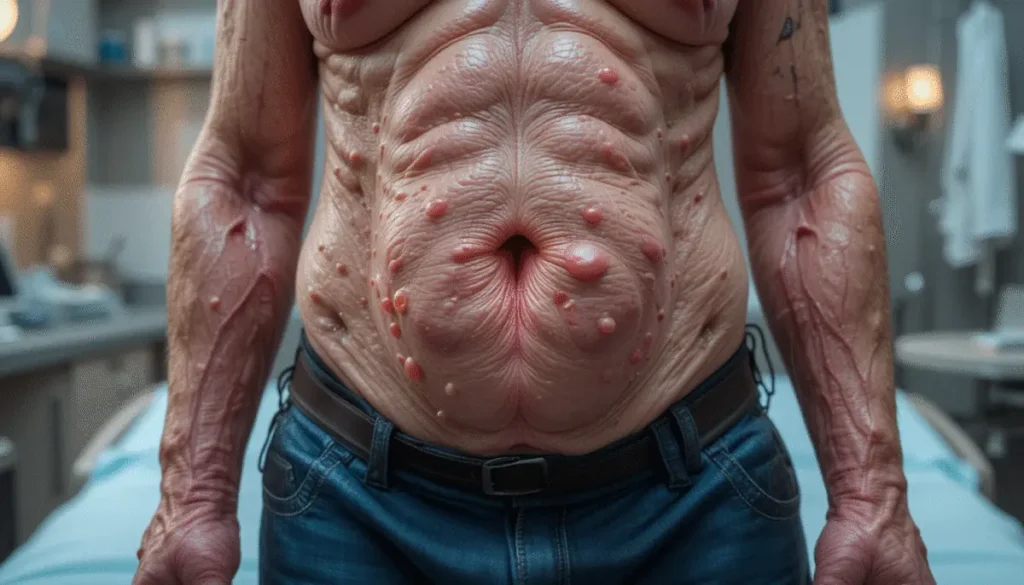
2. Bullous Impetigo
| Feature | Description |
|---|---|
| Cause | Staphylococcus aureus (exfoliative toxin) |
| Lesions | Flaccid, fluid-filled blisters (bullae) |
| Location | Trunk, limbs |
| Symptoms | May be more rapidly spreading |
| Lymphadenopathy | Less common |
📸 Yellow, fluid-filled bullae that enlarge rapidly and rupture to leave shallow erosions.
💊 Treatment of Impetigo
✅ Step 1: Crust Removal
- Soak affected area with wet gauze or warm compress to remove crusts
- This enhances topical antibiotic penetration
✅ Step 2: Topical Antibiotics (for localized disease)
| Medication | Directions |
|---|---|
| Mupirocin 2% ointment | Apply 3× daily for 7–10 days |
| Fusidic acid (if available) | Alternative in some settings |
✅ Step 3: Oral Antibiotics (for widespread/extensive disease)
| Medication | Dose & Duration |
|---|---|
| Dicloxacillin | 7–10 days |
| Cephalexin (1st-gen cephalosporin) | Preferred for broader coverage |
| Clindamycin or Erythromycin | For penicillin-allergic patients |
🩺 Prognosis
- Excellent prognosis with treatment
- No scarring as infection is superficial
- Recurrence possible in unhygienic or crowded settings
- Encourage hygiene education to reduce spread
📝 Summary Table
| Category | Details |
|---|---|
| Disease | Impetigo – superficial bacterial skin infection |
| Affected Layer | Epidermis |
| Common in | Children, warm climates, poor hygiene |
| Non-Bullous Type | Honey-crusted lesions – Streptococcus ± Staph |
| Bullous Type | Fluid-filled bullae – Staphylococcus aureus |
| Treatment (mild) | Mupirocin ointment after crust removal |
| Treatment (severe) | Oral dicloxacillin, cephalexin, or alternatives |
| Prognosis | Heals well, no scarring, recurrence possible |